
Catalog of Regulatory Science Tools to Help Assess New Medical Devices
This regulatory science tool (RST) is a software framework for generating hybrid breast CT images that combine simulated microcalcification clusters with real patient breast CT projection data.
Technical Description
This tool implements a hybrid virtual imaging trial (VIT) framework for breast computed tomography (CT) by combining simulated microcalcification clusters with real patient breast CT projection data. The framework performs lesion insertion in the projection domain, allowing simulated pathology to undergo the same physical image formation processes as the anatomical background, including polyenergetic attenuation, detector response, geometric effects, and image reconstruction.
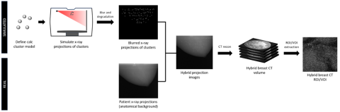
Simulated microcalcification clusters are generated using configurable parameters (size, number, composition, and density) and forward-projected using GPU-accelerated ray tracing. The simulated projection data are combined with patient projection data prior to reconstruction to generate hybrid projection images. The hybrid datasets are then reconstructed using user-defined reconstruction algorithms and parameters to produce volumetric breast CT images suitable for task-based evaluation. An overview of the hybrid simulation workflow is shown in Figure 1.
Tool Components: Python simulation scripts, sample system-specific configuration files (geometry, spectra, energy spectra, materials, MTF)
Tool Inputs: The tool requires:
- Patient breast CT projection images
- Patient segmentation volumes, labeled as air, adipose, fibroglandular tissue, and skin
- X-ray energy spectrum for the breast CT system
- System geometry parameters
- Detector modulation transfer function (MTF)
- User-defined microcalcification parameters
Tool Outputs: The tool generates:
- Hybrid log-normalized projection datasets
- Reconstructed hybrid breast CT volumes
- Extracted signal-present and signal-absent volumes of interest (VOIs)
- Maximum intensity projection (MIP) images
Intended Purpose
The hybrid simulation framework is intended to generate virtual breast CT images by inserting simulated microcalcification clusters into real patient projection data. The tool reproduces key components of the X-ray imaging chain, including energy-dependent attenuation, detector response, geometric effects, and image reconstruction, to produce anatomically realistic hybrid datasets. The framework is intended for task-based evaluation of microcalcification detectability in dedicated breast CT systems. The generated datasets can be used to study the impact of lesion characteristics, reconstruction methods, and system parameters on detection performance using human observers or model observers.
This tool may be used in device research and development, regulatory science investigations, and methodological studies involving breast CT imaging performance. The modular design allows implementation with user-supplied patient datasets and system-specific parameters.
Testing
Validation of the hybrid simulation approach is presented in this journal article [Ref 1]:
S. H. Lyu, A. Makeev, D. Li, A. Badal, A. M. Hernandez, J. M. Boone, and S. J. Glick, “Hybrid simulation of breast CT for assessing microcalcification detectability,” Journal of Medical Imaging, vol. 12, Jul. 2025, doi: 10.1117/1.JMI.12.S2.S22015.
This simulation framework was validated using real patient breast CT datasets obtained under a Material Transfer Agreement (MTA) from the University of California, Davis [Ref 2]. The results demonstrate that the approach can quantify detectability performance under controlled variations of lesion and system parameters.
The tool is designed to be adaptable; users may implement the framework with their own patient datasets and system-specific parameters to evaluate imaging performance for their respective breast CT platforms.
Limitations
The tool requires access to patient projection data and segmentation volumes. The simulation framework does not currently model:
- Scatter originating within calcifications
- Quantum noise specific to simulated lesions
- Spatially variant MTF effects
Supporting Documentation
GitHub repository: https://github.com/DIDSR/hybrid-breastCT
Related Work:
[1] S. H. Lyu, A. Makeev, D. Li, A. Badal, A. M. Hernandez, J. M. Boone, and S. J. Glick, “Hybrid simulation of breast CT for assessing microcalcification detectability,” Journal of Medical Imaging, vol. 12, Jul. 2025, doi: 10.1117/1.JMI.12.S2.S22015.
[2] P. M. Gazi et al., “Evolution of spatial resolution in breast CT at UC Davis,” Med. Phys. 42(4), 1973–1981 (2015).
Contact
Tool Reference
- RST Reference Number: RST26MD04.01
- Date of Publication: 05/04/2026
- Recommended Citation: U.S. Food and Drug Administration. (2026). Hybrid Simulation Framework for Breast CT Virtual Imaging Trials (RST26MD04.01). https://cdrh-rst.fda.gov/hybrid-simulation-framework-breast-ct-virtual-imaging-trials